animal cell under microscope labeled
They are different from plant cells in that they do contain cell walls and chloroplast. All information about animal cell under microscope labeled.

Cell Organelles And Their Function
Ease of use and automation from a fully integrated microscope platform and.

. 5th Grade Science Worksheets Cells -. It also shows the myoepithelial cells that surround each sweat gland of the animal skin. These regions of growth are good for studying the cell cycle because at any given time you can find cells that are undergoing mitosis.
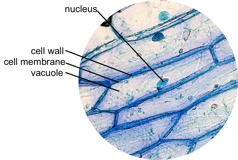
Onion Cell drawing high power 2. Animal Cell- Definition Structure Parts Functions Labeled Diagram Amazing 27 Things Under The Microscope With Diagrams Prokaryotes vs Eukaryotes- Definition 47 Differences Structure Examples. Here in the diagram you will see some seminiferous tubules lined by the.
You will find two main parts in hair a cylindrical shaft and a terminal hair follicle. Animal Cell Under Microscope Labeled Cell Structure - There are three structural parts of the microscope ie. Plant and Animal Cells Microscope Lab Author.
Get more skin-labeled diagrams on social media for anatomy learners. Within the epidermis of a skin you will find squamous diamond-shaped and polyhedral cells under the light microscope. Animal cells are eukaryotic cells that contain a membrane-bound nucleus.
Cells are the basic unit of life and these microscopic structures work together and perform all the necessary functions to keep an animal alive. Most of the cells size range between 1 and 100 micrometers and are visible only with the microscope. Most cells both animal and plant range in size between 1 and 100 micrometers and are thus visible only with the aid of a microscope.
The structure of an animal cell with labeled parts. A cell is the smallest functional and structural entity of life that it is easier observing animal cell under light microscope lensclutcolunch. Skin cells under a microscope.
Gently swirl the contents to cover the cell layer. Hair under microscope. The space inside of the cell is known as the cytoplasm.
Plant cells have cell walls one large vacuole per cell and chloroplasts while animal cells will have a cell membrane only. Cheek Cell Lab Gracyns Blog - You get the best of both worlds. I will show you the sperm under a microscope 400x with the labeled diagram.
Under the microscope animal cells appear different based on the type of the cell. So hair is an epidermal down growth embedded into the dermis or hypodermis of the animals skin. Examining animal cells under the microscope.
Animal Cell Under A Microscope Labeled Cell-fie. Here the seminiferous tubules of the animal show different types of cells like primary spermatocytes secondary spermatocytes spermatid and spermatozoa. Students will observe onion cells under a microscope.
These are both specific typesof cells. Simple columnar epithelium under a microscope The simple columnar epithelium under a microscope consists of a single layer of taller than wide cells. Labeled animal cell below electron.
Learn the structure of animal cell and plant cell under light microscope. Neuron under microscope labelled diagram. These simple columnar epithelia look closely packed and slender columns shaped.
Labeled animal cell under electron microscope 8745961 orig. The animal cell diagram is widely asked in Class 10 and 12 examinations and is beneficial to understand the structure and functions of an animal. A brief explanation of the different.
Failure to cluster the parental genomes leads to chromosome segregation errors and micronuclei which are incompatible with healthy embryo development. Animal cell microscope labeled. All organisms are made up of cells or in some cases a single cell.
The cylindrical shaft of the hair under a microscope shows three layers medulla cortex and cuticle of keratinized cells. A typical animal cell is 1020 μm in diameter which is about one-fifth the size of the smallest particle. The large spherical area is the nucleus while the granulated part is the cytoplasm of the cell.
Animal cells have a basic structure. You should observe the cell membrane nucleus and cytoplasm. You will find the cell base on the basement membrane and the apex in contact with the lumen.
Draw a diagram of one cheek cell and label the parts. The Cell Structure Gizmo allows you to look at typical animal and plant cells under a microscope. Technology Last modified by.
A cell is the smallest functional and structural entity of life that it is easier observing animal cell under light microscope. Animal Cell Diagram Under Microscope Labeled. A cell is the smallest functional and structural entity of life that it is easier observing.
Electron cell microscope animal labeled under labels label orig using structures identifying function use. Coli cell which is a type of bacteria. Sperm under microscope 400x labeled.
You perform superresolution live cell imaging with up to 15 resolution improvement. As you can see in the above labeled plant cell diagram under light microscope there are 13 parts namely Cell membrane.

Animal Cell Structure And Organelles With Their Functions

Eukaryotic Cell Structure Animal Cell Plant And Animal Cells Eukaryotic Cell

Diagram Of Plant And Animal Cell Under Electron Microscope

Visualization Of The Cell Using Em

Cell Nucleus Function Structure And Under A Microscope

Cells And Dna Lesson Plan Science Cells Middle School Science Activities Dna Lesson Plans

Printable Labeled And Unlabeled Animal Cell Diagrams With List Of Parts And Definitions Cell Diagram Animal Cell Animal Cells Model

Explore Collection Of Muppets Animal Drawing

Lab The Cell The Biology Primer

Science Archives My Teaching Library Chsh Teach Llc


